Set up
- Probe: Linear
- Mode: Peds Abdomen
- Starting Depth: 5 cm
Views
- No intussusception visualized – Clip of each sweep (ascending, transverse, and descending colon)
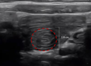
- Intussception visualized
- Short axis with measurement
- Long axis
Scanning
- Place the patient in supine position
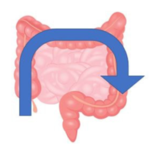
- Start with the probe marker to the patient’s right in the RLQ and scan along colon as indicated in the figure below keeping probe perpendicular to the colon
- Use graded compression to move gas out of view to visualize the bowel
- Search for a non-compressible tissue structure along this path
Abnormal Measurements
- > 2.5 cm
RLQ landmarks

RUQ landmarks
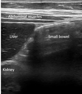
Normal bowel
Colon and small bowel

Transverse view “target sign”
Long axis view “sandwich sign”
Ileo-Ilial Intussusception
< 2cm, usually with peristalsis
Intussusception mistaken for a lymph node or kidney.
Fix → Intussusception can be distinguished from lymph node by size and presence of concentric circles (loops of bowel).Identify the kidneys separately to distinguish from intussusception.
Small bowel intussusception can look similar to ileocolic intussusception
Fix → Measure abnormal structure, if less than 2cm, is small bowel intussusception and does not get referred for enema reduction.