Set-up
- Probe: Phased array
- Mode: Peds Cardiac
- Starting Depth: 10 cm
Views
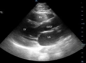
- Parasternal long axis
- Parasternal short axis
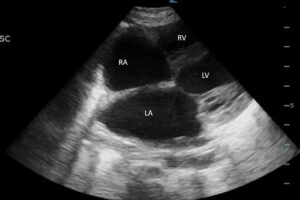
- Apical 4-view
- Subcostal
- IVC
Scanning
- PSL- probe marker toward patient’s right shoulder at the 4th intercostal space just left of sternum
- PSS-Rotate 90 degrees with probe marker to patient’s left shoulder
- A4-Slide probe down to apex and lower handle toward patient with maker facing 3 o’clock position (should be about nipple line)
- SC-Move probe under xiphoid with indicator to patient’s left, handle against abdomen and probe directed to left chest to obtain subcostal view
- IVC-indicator toward patients feet, handle at 90 degrees to bed, rock probe to look up and visualize IVC at juncture with RA
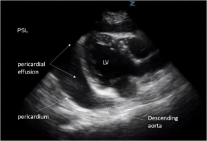
Parasternal Long Axis
Parasternal Short Axis

Apical 4
Assess fractional shortening
RV/LV ratio
Mitral/Tricuspid valves

Subcostal

IVC

Pericardial Effusion
Fluid present between heart and pericardium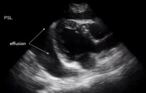
Dilation of atria and ventricles
Dilated right ventricle

Cardiac tamponade
Pericardial fluid + RV collapse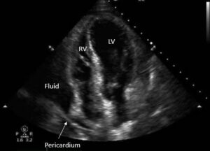
Pleural effusion mistaken for pericardial effusion
Fix → Pericardial effusions taper anterior to the descending aorta, whereas pleural effusion extend posterior
Fix → Visualize the IVC entering the right atrium, and locate aorta to left of IVC in abdomen
